Posts

Showing posts with the label Cell

MINISTRY OF DEFENCE

Image
  MINISTRY OF DEFENCE. Establishment Of National University In Islamabad Total position. 29 Domicile , All over Pakistan    Qualification  intermediate to bachelor positions . Project Engineer(Civil) account Officer  supervisor assistant office boy     How to apply.   Apply online through the link https://www.fgei-cg.gov.pk/nup-rec.php   Last date to apply  02 April 2023. No TA/DA Fo Interview 

Endoplasmic Reticulum (Structure and Function)

Image
Endoplasmic Reticulum (Structure and Function). Endoplasmic reticulum  It is a network of channels present throughout the cell most of these channels are continuous with the plasma membrane toward the outer side on their inner side. They are in contact with the nuclear membrane.   The entire system of channel is called endoplasmic reticulum.  Morphologically  Endoplasmic reticulum occurs in three forms. 1. Cisternae:  The channels of Endoplasmic Reticulum are composed of spherical or tubular membranes are called cisternae. (pancreas, notochord,brain).  2. Vesicles the vesicle are oval membrane-bounded vascular structure having the diameter of 25 to 500 micrometre.  3.Tubules The tubules are branched structure forming the reticular system along with the cisternae and vesicle. Types of Endoplasmic Reticulum:   There are two types  1. Granular or rough endoplasmic reticulum.    Ri...

Lysosomes (Structure and Function)

Image
Lysosomes (Structure and Function) ( Lysosomes in Urdu) lysosomes The word lysosome is taken from Greek word (Lyso= digestive,splitting+ soma=body) so, they are concerned with breaking activities. These are present in most animals and few plants cell . They are absent in bacteria and mature mammalian erythrocyte. Structure Lysosome are 0.5 to 0.2 micro metre in size. They are bounded by a single membrane, this membrane forms a simple check and  this sac contains several hydrolytic enzymes like acid phosphatase. According to a recent estimate a lysosome contain up to 40 types of hydrolytic enzymes. All lysosomal enzymes are acid hydrolysis optimally active at the pH 5 maintained within lysosomes. Formation They are formed by Golgi bodies. Kinds of Lysosomes Morphologically there are four types.  The first is primary lysosome and other three (heterophagosome, autophagosome And residual bodies) have been grouped together as secondary lysosome. Disease...

Cell Wall

Image
Cell Wall The outermost boundary in most of the plant cell is cell wall. It is present in plants, Fungi, bacteria and absent in animals. Cell walls have different structures and chemical compositions. Cell wall is non living and secreted by the protoplasm . Formation of cell wall takes place at telophase of cell division.        In a young cell, the cell wall is thin and delicate, but in large cell, it becomes thick and strong. The cell wall consist of three layers. i) Primary Wall It is found around a young plant cell on the outer side of Cell Membrane. The primary wall is very thin, elastic and capable of great extension. It consist of cellulose l, hemicellulose, pectin and sugar compounds. The cellulose molecules are arranged in a criss cross manner. ii) Secondary Wall: It is formed on the inner surface of the primary wall. It is much thick and rigid. It is composed of inorganic salts, silica, waxes, cutin, lignin, etc iii) Middle Lamel...

Centrosome and Golgi Bodies

Image
Golgi Bodies And Centrosome Golgi bodies Golgi bodies were discovered by  camelo Golgi in 1898.  These are present in animal and also in plant cells.  The Golgi bodies are composed of phospholipid and protein, they contain 3 to 20 sacs. The  Golgi apparatus  is a collection of membrane enclosed in sacs (cisterns)  that are  stalked like dinner plates. These cisternae are associated with vesicle  called Golgi vesicles. The Golgi complex is a complex system of interconnected tubules,  these tubules are present around Central stocks. One or more Golgi apparatus are present in all Eukaryotic cells.  Golgi is directed at proper glycosylation of protein and lipids.  There are more than 200 enzymes that function to add remove are modified Sugars from proteins and lipids in the Golgi apparatus.  The Golgi apparatus in polarized structure with  cis and trans sides. Newly synthesized proteins bud off from the gr...

Molecular motors (kinesin,,Myosin,Dynesin)

Image
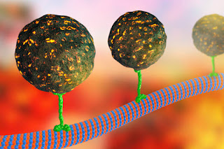
Molecular Motors Molecular motors :molecular motors that move proteins,organelles and other cell parts ( collectively cargo )to all parts of the cell are  100 to 500 KDA ATPases .  ==> Attach to their cargo at one end of the molecules and two microtubules are actin Polymers with the other end had. ==> Convert the energy of ATP into movement along the cytoskeleton . three super families of molecular motors Kinesin  It  is double headed molecule that moves its cargo towards the positive ends of microtubules and then bends its neck to other head. ==> Some can kinesin are associated with mitosis and meiosis. ==> Other kinesin perform different function movement Kargo to the negative end of microtubule . 2 Myosin  ==> Divided into 18 classes.  == The heads of the myosin bind to Actin and produce motion by bending the neck  regions or walking along microfilament one head after the other 3 Dynesin  ==> Have ...

Nucleus

Image
Nucleus Robert BrownThe title of the discovery of the Nucleus goes to Robert Brown in 1831, it looks dark than the surrounding cytoplasm. the shape of nucleus may be spherical or irregular. it controls the life and activities of the cell,in animal it is present in centre of cell but in plant it is pushed towards a side due to large vacuole. the number of nucleus is different in different cell. mononucleate the cell have one nucleus.   Binoculeate :  Cell have two nuclei.   Multinucleate : The cell with more than two nuclei. The nucleus is visible only during the non dividing phase. Chemically the nucleus is composed of DNA,RNA and protein including enzymes.  nucleus is composed of four  parts 1 Nuclear membrane:  it separates the nuclear material from the cytoplasm,it is not a complete barrier, It contains pores called nuclear pores.over 3,000 nuclear pores are present in egg but erythrocyte have  only 3 to 4 pores. Certain sub...

Cytoskeleton,(Microtubules,Intermediate Filament,Microfilament

Image
Cytoskeleton (Microtubules,Intermediate Filament,Microfilament Cytoskeleton : skeleton of cell maintain the structure of cell.  permits the cell to change shape and move. Cytoskeleton composed of microtubules,microfilaments and intermediate filaments. microtubules they are long  unbranched and slender structure. ==> Road inside cell for transportation.  Made up of two Globular protein,Alpha tubulin and  beta tubulin,they are involved in the formation and degeneration of spindle during cell division. = = > Several cell organelle are derived from microtubules,these organelles are cilia,flagella,basal bodies and Centriole. ==> They provide the tracks along with several different molecular moters move,transport vesicle, organelles such as secretary granules and mitochondria . ==> The anticancer paclitaxel  (taxol) binds to microtubules and make them so stable so that organelles can't move, mitotic spindle can't form and the cel...

Mitochondria

Image
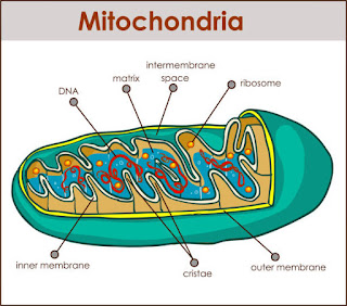
Mitochondria   Mitochondria are very important organelles they are present only in Eukaryotic cell they are involved in manufacturing and supply of energy to the cell.mitochondria have the ability to form the energy rich compound ATP by oxidative phosphorylation.  mitochondria called powerhouse of cell, mitochondria are the centre of aerobic respiration. The size and number of Mitochondria is different in different cells. it is about 0.2 to 1.0 nanometre in diameter and about 10 micrometre long. each Eukaryotic cell may have hundreds to thousands  of Mitochondria. Structure Of Mitochondria: mitochondria are double membrane bounded organelles,the outer membrane is smooth and the inner membrane forms many infolding called Cristae . the inner Chamber of Mitochondria is called mitochondrial matrix ,the inner surface of cristae contains small Nobe like structure called F1 particles ,these particle are suspended inside the matrix. the Cristae increase the surf...

Plasma Membrane/Plasma Lemma

Image
Plasma Membrane Plasma Membrane: Outer most layer in animal cell and second outer most layer in plant cell. It is also called plasma-lemma. Structure of plasma membrane was proposed by two scientists Singer and Nicolson in 1972,called fluid Mosaic Model. According to this model plasma membrane is composed of two layer of phospholipid,(phosphatidylcholine and phosphotidylethanolamine). Along these lipids proteins are also present. Each layer of phospholipid has two ends. 1. Hydrophobic end  uncharged tail end insoluble,it is nonpolar end, inner part these ends are present opposite  to each other. 2. hydrophilic end polar end (outer part) has charged,head end of the molecule contains  phosphate  portion and it is is relatively soluble in water. in these layers proteins are present in different manner. 1. extrinsic proteins  also called peripheral proteins these proteins are present along the surface of lipids they have loose attachment wi...

Popular posts from this blog

What are chromosomes?

Neurotransmitters and receptors

Excretion in Invertibrate,(Excretion in Planaria, Excretion in Earthworm,Excretion in Cockroach)